


Retina Associates, a Tucson, Arizona-based ophthalmology practice in operation for more than 50 years, specializes in diagnosing and treating diseases affecting the retina, macula, and vitreous, and its physicians are board certified through the American Board of Ophthalmology. The practice is led by chief physician Cameron Javid, MD, who completed his ophthalmology residency at Tulane University Medical Center and his retina fellowship at the Massachusetts Eye and Ear Infirmary/Harvard Medical School, and who also serves as a clinical associate professor of ophthalmology at the University of Arizona College of Medicine. Retina Associates has participated in numerous national clinical research trials examining conditions such as macular degeneration, including studies involving gene therapy for retinal conditions and late-stage dry age-related macular degeneration, an area closely tied to ongoing research into how lesion growth is measured in clinical trials for geographic atrophy.
Why Lesion Growth Guides Geographic Atrophy Trials
Geographic atrophy can expand while a person still retains useful central vision. That mismatch creates a difficult research problem: A study cannot rely only on a vision chart to detect whether an investigational approach has changed the course of the condition. Researchers therefore need an outcome that can register structural change before every consequence is apparent in daily tasks.
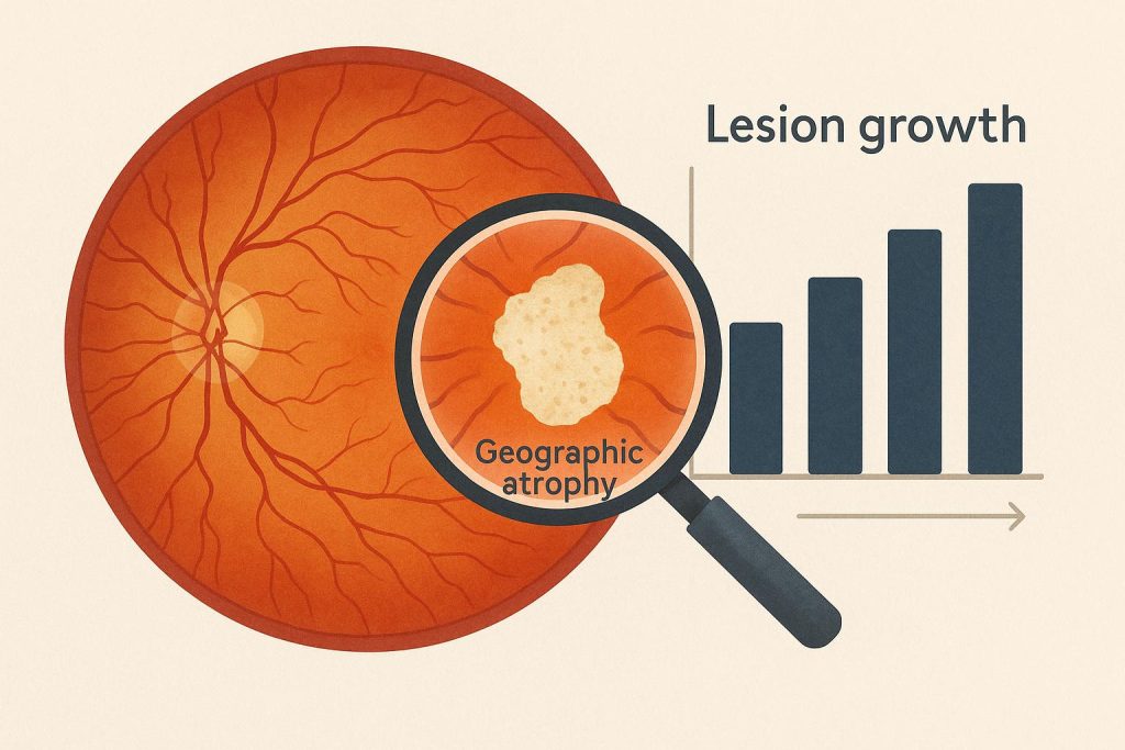
The purpose is to detect patterns that can be compared between study groups over time. That need has made lesion growth a central measure in trials involving geographic atrophy, an advanced form of dry age-related macular degeneration. In this setting, a lesion is an area of retinal damage.
Tracking its size over time does not turn an image into a prediction for any one person. It does, however, give a trial a defined way to compare the pace of change between groups.
Fundus autofluorescence is one tool used for that comparison. The imaging method records natural light signals from the back of the eye, helping trained readers outline affected areas on repeat images. Once a study uses the same imaging approach at planned intervals, investigators can calculate a growth rate rather than relying on a single photograph. Consistency matters because the endpoint depends on comparing like with like across the study period.
The pivotal studies of avacincaptad pegol, later approved as Izervay, illustrate the logic. Their primary endpoint compared the average rate of lesion expansion from baseline through month 12. Images were assessed at baseline, month 6, and month 12, which allowed the analysis to focus on a slope of change instead of a simple before-and-after difference.
That distinction matters because geographic atrophy does not progress in identical patterns. A larger lesion at enrollment may have a different starting point from a smaller one, while a vision score can be influenced by where damage lies within the macula. Measuring the rate of expansion gives researchers a common frame for evaluating groups, even when individual experience remains more complex than one endpoint. For this reason, protocols define when images are collected and how those images are evaluated. The approach also makes it possible to examine whether a difference persists over a defined follow-up period.
The endpoint also clarifies what a trial result does and does not mean. A slower average growth rate describes a difference in structural progression within the studied population. It does not establish that every participant will experience the same change, nor does it capture every aspect of visual function. Trial reports should be read alongside the study design, follow-up period, participant characteristics, and safety findings.
For clinicians and research teams, that discipline affects communication as much as analysis. A discussion of lesion growth can explain why follow-up imaging matters without converting a population measure into a personal forecast. It can also distinguish an approved use from an investigational question, a useful boundary as studies continue to examine new approaches for retinal disease.
The broader point is practical: A strong clinical trial endpoint matches the feature of disease that a study is designed to track. In geographic atrophy, repeatable measurement of lesion expansion offers a way to test whether the pace of anatomical change differs over time. That measurement does not replace functional assessment, but it supplies a consistent foundation for interpreting a trial’s central finding.
About Retina Associates
Retina Associates is a Tucson, Arizona ophthalmology practice that has served patients for more than 50 years, specializing in the diagnosis and treatment of diseases affecting the retina, macula, and vitreous. Its physicians are board certified through the American Board of Ophthalmology, and the practice is led by chief physician Cameron Javid, MD, who also serves as a clinical associate professor at the University of Arizona College of Medicine. The practice has participated in numerous national clinical trials focused on macular degeneration.